|
Singapore-MIT Alliance for Research & Technology |
BioSystems and Micromechanics (BioSyM) Inter-Disciplinary Research Group |
||||||||||
|
|
BioSyM ResearchThrust 4: In vivo cell systems analysisResearch Projects
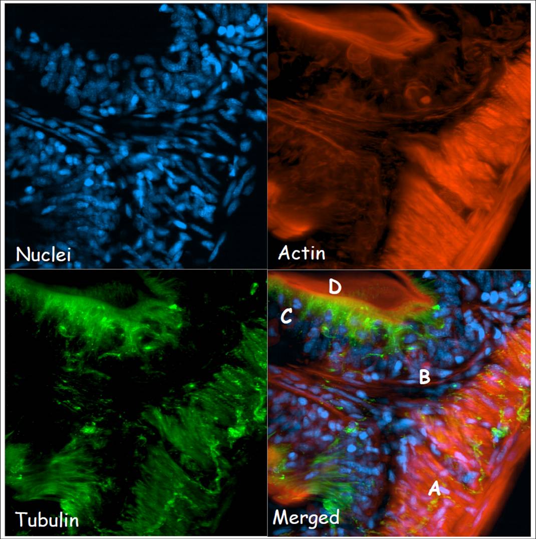
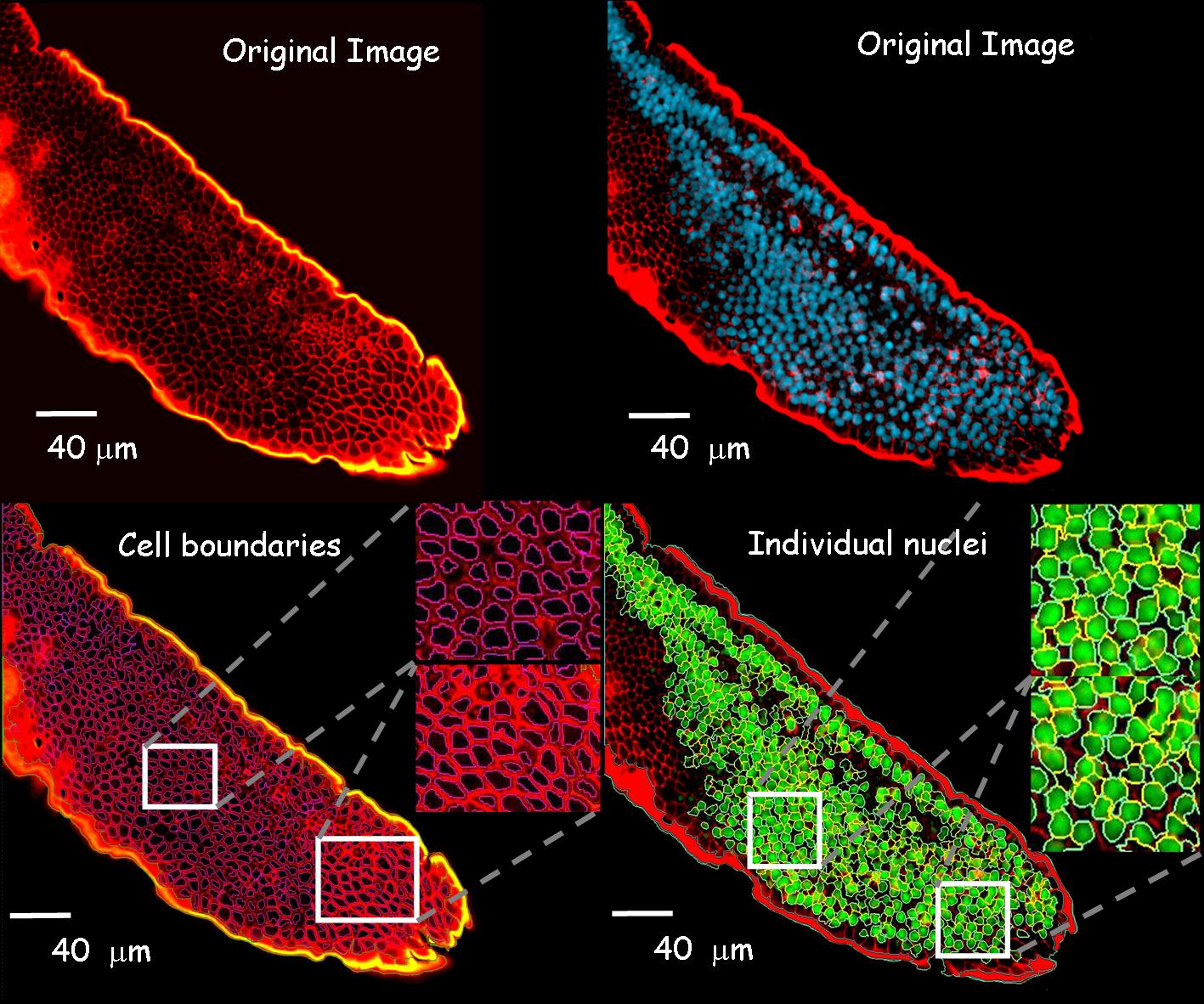
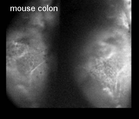
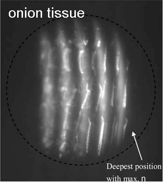
1. Novel optical microscopy and phase recovery methods for bioimaging(George Barbastathis (MIT/SMART-BioSyM), Peter T. C. So (MIT/SMART-BioSyM), Roger D. Kamm (MIT/SMART-BioSyM), H. Harry Asada (MIT/SMART-BioSyM), Paul Matsudaira (NUS), Colin J. R. Sheppard (NUS), Dipanjan Battacharya (SMART-BioSyM), Se Baek Oh (MIT), Justin Wu Lee (MIT) and Laura Waller (MIT))This project aims to develop non-invasive methods of imaging and phase recovery for imaging cells and tissue in vivo or in micro-biofluidic systems. We have developed optical instruments based on (1) scanning light-sheet microscopy, (2) volume holographic pupil microscopy, (3) colour transport of intensity. These provide three-dimensional image information in real time and with high resolution. Regeneration of intestinal epithelial tissue is a complex dynamic process that involves division and differentiation of stem cells at the base of the villi, migration towards the tip and apoptosis at villi tip. But the basic dynamics of this serial process has not been studied directly. To understand how cells in the intestinal epithelium migrate and the interactions between molecular motors and structural proteins, we are developing a laser sheet based microscope and other novel optical methods such as Multiplex-Volume holographic microscope to achieve improved spatio-temporal resolution. 1. Development of Laser-sheet based microscope for Bioimaging: We have built a SPIM (Selective Plane Illumination Microscopy) for live small animal imaging and are working on developing microscopic techniques towards live small animal imaging. In preliminary studies we are imaging with standard confocal approaches fixed intestinal tissue of mice, zebrafish and medaka fish model system to understand organization of different structural proteins forming intestine. Although confocal microscopy provides us a good x-y spatial resolution, but z-axis and temporal resolution are very poor. Also, confocal microscope has high photo-bleaching effect, not suitable for live small animal imaging. Using laser sheet based microscope we can excite a thin section of tissue (~micron) and collect the emission of this complete fluorescent plane simultaneously in a cooled CCD. We have built a laser sheet-based microscope and are working to further development of this technique for higher spatial resolution.
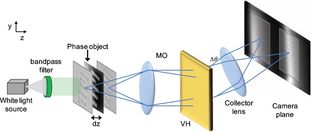
2. Development of Multiplex-Volume holographic microscope: We have started to build Multiplex volume hologram (MVH) microscope for spatial-spectral 4D imaging systems for real time imaging. In this method using a small multiplex volume holographic material in the collection pathway, we can spatially separate fluorescent emission from different fluorescent planes and simultaneously acquire spectral images of these planes in different parts of a cooled-CCD simultaneously. Using this method we can capture 3-D images of a specimen, simultaneously, without any scanning.
2. Transformation optics for cellular imaging and manipulation(Baile Zhang (SMART-BioSyM), George Barbastathis (MIT / SMART-BioSyM), Collin Sheppard (NUS), Hanhong Gao (MIT) and Satoshi Takahashi (MIT))Biological imaging at the molecular and cellular scales requires resolving of features in the order of 10's of nanometers. However, the fundamental diffraction limit discovered by Abbe in 1873 restricts the resolution of a conventional optical microscope to about half a wavelength (about 200 nm in air) due to the loss of high spatial frequency information carried in the evanescent waves. It is strongly desirable to develop a real time imaging technique that can provide subwavelength information. We propose to use gradient index (GRIN) dielectric materials to design and manufacture practical devices that can significantly improve the resolution of a common far-field microscope. Current simulations have shown that it is possible to achieve sub-100 nm resolution over broadband of visible spectrum by adopting this kind of device. Different from previous superlens and hyperlens that utilize surface plasmon resonance between metal and dielectric to capture subwavelength features within narrowband, our approach has great advantages (low-loss and broadband) and will be greatly simplified with existing micro-manufacturing technologies.
|
|
|||||||||